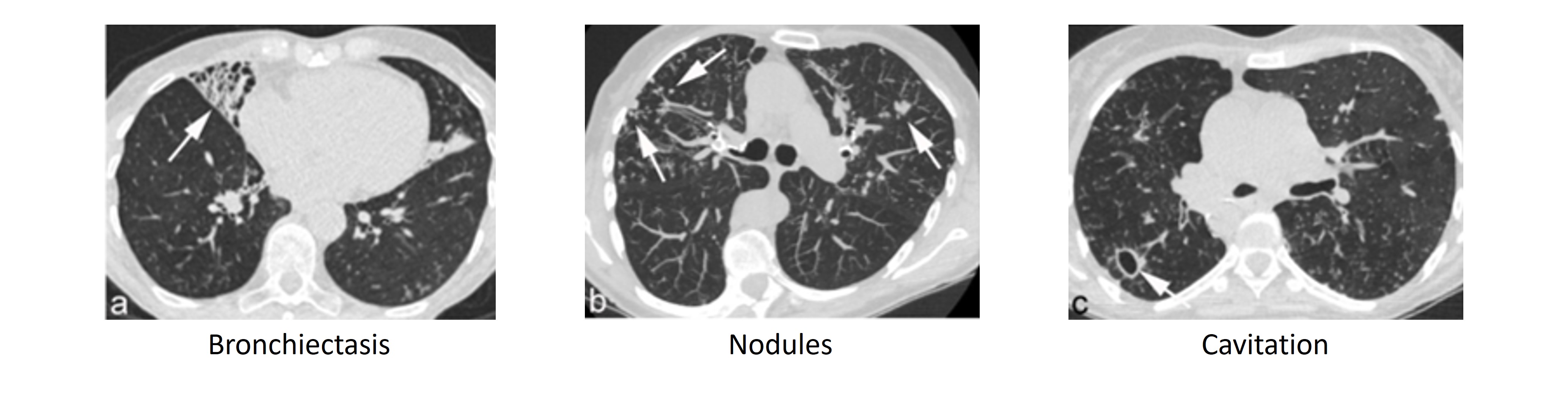
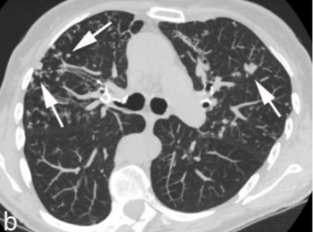
Symptoms and Diagnosis

  • The diagnosis of nontuberculous mycobacteria (NTM) pulmonary disease relies on the combination of clinical, radiographic, and microbiologic criteria1
  • Symptoms of NTM pulmonary disease are variable and non-specific; however, a chronic productive cough is present in most patients2-4
  • Positive cultures from respiratory clinical samples and characteristic radiographic findings are also required to confirm the diagnosis of NTM pulmonary disease2
Image
Symptoms and diagnosis

The Diagnosis of NTM Pulmonary Disease

ATS/IDSA Diagnostic Criteria for NTM Pulmonary Disease1


Image

Clinical Features of NTM Pulmonary Disease

The symptoms of NTM pulmonary disease are variable and nonspecific, which may result in a delayed diagnosis.2,3 Most patients with NTM pulmonary disease have chronic or recurring productive cough.2 Constitutional symptoms may also be present, especially in the setting of more advanced disease.2 Symptoms should be monitored regularly as worsening may indicate disease progression.2,5 

Radiographic Features of NTM Pulmonary Disease

Lung radiographs help to establish the initial NTM pulmonary disease diagnosis as well as monitor disease progression over time. 


On a high-resolution computed tomography scan, NTM pulmonary disease may be observed as bronchiectasis with multiple small nodules. Nodular or cavitary opacities can be seen on a chest radiograph.
 

Radiographic Computed Tomography Findings in Patients with NTM Pulmonary Disease

Image

Images from Dettmer S, et al. J Clin Med. 2021;10(12):2736 under the Creative Commons Attribution License (CC BY).6

Two distinct radiographic presentations of NTM pulmonary disease are typically observed — nodular-bronchiectatic or fibrocavitary — each with their own common patient characteristics.2,5

Nodular-bronchiectatic Disease3,5


Nodular-bronchiectatic disease often progresses slowly over many years. 

Typical patient characteristics include: 
 

  • Women
  • Older age
  • Nonsmokers
  • Tall and thin with low body mass index
Image

Computed Tomography Scan Showing Lung Nodules

Computed tomography scan reveals multiple lung nodules indicated by the white arrows.  
Image from Dettmer S, et al. J Clin Med. 2021;10(12):2736 under the Creative Commons Attribution License (CC BY).6
 

Fibrocavitary Disease5


Fibrocavitary disease may have more rapid disease progression.
Typical patient characteristics include: 


  • Male
  • Older
  • Smokers
  • Various body builds
Image

Chest Radiograph Showing Multiple Cavities

Chest radiograph shows parenchymal opacity which contains multiple cavities in the right upper lobe. Image is unmodified from Koh WJ, et al. J Korean Med Sci. 2005;20:913 under the Creative Commons Attribution Non-Commercial License (CC BY-NC).7

Microbiologic Features of NTM Pulmonary Disease

The isolation of NTM from respiratory clinical samples is essential for the diagnosis of NTM pulmonary disease.2 Once antimycobacterial therapy is initiated, sputum cultures are also followed serially over time as a marker of treatment response.


Microbiologic findings consistent with NTM pulmonary disease include: 


  • NTM positive cultures from sputum, bronchoalveolar lavage fluid, or tissue specimens1,2 
  • Two or more positive cultures with the same NTM species, if sputum cultures are used
  • All cultures for NTM should include both solid and broth (liquid) media for the detection and enhancement of growth2

Solid media 

Solid media allow for the observation of colony morphology, growth rates, recognition of mixed mycobacteria infections, and quantitation of the infecting organism.2


Broth Media

Cultures in broth media have a higher yield of mycobacteria and produce more rapid results than those on solid media, but may not be satisfactory on their own because of bacterial overgrowth.


Image

Example of Colonies on Solid Media

Image is unmodified from Gieger A, et al. Nepal J Ophthalmol. 2017;9(1):91–4  under the terms of the Creative Commons Attribution Non-Commercial No Derivatives License (CC BY-NC-ND).8  

References

  1. Daley CL, Iaccarino JM, Lange C, et al. Treatment of Nontuberculous Mycobacterial Pulmonary Disease: An Official ATS/ERS/ESCMID/IDSA Clinical Practice Guideline. Clin Infect Dis. 2020;71(4):e1-e36. doi:10.1093/cid/ciaa241
  2. Griffith DE, Aksamit T, Brown-Elliott BA, et al. An official ATS/IDSA statement: diagnosis, treatment, and prevention of nontuberculous mycobacterial diseases. Am J Respir Crit Care Med. 2007;175(4):367–416. doi:10.1164/rccm.200604-571ST
  3. Kim RD, Greenberg DE, Ehrmantraut ME, et al. Pulmonary nontuberculous mycobacterial disease: prospective study of a distinct preexisting syndrome. Am J Respir Crit Care Med. 2008;178(10):1066–74. doi:10.1164/rccm.200805-686OC
  4. Huang JH, Kao PN, Adi V, Ruoss SJ. Mycobacterium avium-intracellulare pulmonary infection in HIV-negative patients without preexisting lung disease: diagnostic and management limitations. Chest. 1999;115(4):1033–40. doi:10.1378/chest.115.4.1033
  5. Varley CD, Winthrop KL. Nontuberculous Mycobacteria: Diagnosis and Therapy. Clin Chest Med. 2022;43(1):89–98. doi:10.1016/j.ccm.2021.11.007
  6. Dettmer S, Ringshausen FC, Fuge J, et al. Computed Tomography in Adults with Bronchiectasis and Nontuberculous Mycobacterial Pulmonary Disease: Typical Imaging Findings. J Clin Med. 21 2021;10(12)doi:10.3390/jcm10122736
  7. Koh WJ, Kwon OJ, Lee KS. Diagnosis and treatment of nontuberculous mycobacterial pulmonary diseases: a Korean perspective. J Korean Med Sci. 2005;20(6):913-25. doi:10.3346/jkms.2005.20.6.913
  8. Gieger A, Waller S, Pasternak J. Nocardia arthritidis keratitis: case report and review of the literature. Nepalese Journal of Ophthalmology. 2017;9(1):91–94.